17.6.2026Fungal viruses and a 100-million-piece jigsaw puzzle
20.5.2026A cell researcher's new tools
10.4.2026The Mystery of Brain Development
27.2.2026Modelling helps to identify a rare childhood neurological disorder caused by missense mutations of SynGAP1 protein
23.6.2025Too clean is unhealthy
8.5.2025Alkaloids derived from tree bark destroy cancer cells
27.3.2025Vinca alkaloids: Madagascar’s gift to cancer treatment
6.2.2025Genetic testing improves medication safety and effectiveness
26.12.2024The ComPatAI consortium uses large datasets to create an AI learning model for pathology
14.11.2024Microbiota affects the immune system
21.10.2024The skin’s wide range of microbiota improves the immune system
30.9.2024New drug targets from RNA-binding proteins
31.8.2024New machine learning method speeds up drug screening hundred-fold
22.7.2024Mapping the coffee genome to improve disease resistance
25.6.2024Why do some get the severe form of COVID-19?
30.5.2024An AI model that understands health data warns of future diseases
29.4.2024An infrastructure for genomic data
1.4.2024European research community preparing for next pandemic
8.3.2024Evolutionary dynamics of viruses and other microbes affect human health
2.3.2024A million European genomes
20.2.2024Efficient transfer and analysis of biological image data through web interfaces
23.1.2024Improving breast cancer treatment prognoses with liquid biopsy
15.12.2023The European Health Data Space: health data moves across borders for research purposes
16.11.2023New method for measuring gut microbiota
31.10.2023Purifying mining wastewater with plant-associated microbes
29.9.2023Artificial intelligence helps researchers find suitable drugs based on patient’s genetic data and cancer cell samples
1.9.2023Combining data from different sources for personalised treatment
15.8.2023Better treatments for leukaemia
10.6.2023MicroRNAs may reveal type 1 diabetes
16.5.2023Single-cell RNA sequencing enabling individual disease treatment
12.4.2023Tissue samples analysed with Sensitive Data (SD) services provide new information on celiac disease and other autoimmune diseases
20.3.2023DNA isolated from Baltic Sea sediment shedding light on climate change and biodiversity
27.2.2023Organoids grown from stem cells boost cancer research
19.12.2022Sensitive Data (SD) services for Research: with a few clicks a researcher can launch a personal secure computing environment
30.11.2022Microbiota in permafrost play an important role in climate change
20.10.2022Reusable, accurately described and high-quality data – tools created by the research community for agile data management
29.9.2022Gene sequencing used for study of structure and functioning of microbial communities in oceans
1.9.2022Antibiotic-resistant bacteria are a global problem
23.8.2022Personalised medicine against cancer and viruses
30.6.2022Studying the human microbiome is a key towards holistic understanding of our health
23.5.2022FINRISK: one of the world’s longest-running population survey time series
8.4.2022Combining biobank data with data from health registers enables research towards personalised treatment
3.3.2022Finnish research team sequences the genomes of thousands of individuals with diabetes to look for genetic risk factors
10.2.2022BIGPICTURE helps pathology go digital
30.12.2021Sensitive data infrastructure
23.11.2021In the future, an algorithm may diagnose glaucoma from fundus photos
26.10.2021Patient data creating better artificial intelligence models
15.9.2021Teaching an algorithm to identify cancer from sequence data
3.12.2020Efficient processing and sharing of data improving disease diagnosis and treatment
10.11.2020Bioinformatics to revolutionise healthcare: Efficient data processing speeds up diagnoses and enables personalised drug treatments
27.8.2020Tissue samples into digital images, interpreted by artificial intelligence
9.6.2020Digital pathology speeds up diagnosis
18.5.2020Searching markers for breast cancer by machine learning
8.4.2020Metabolomics measures and analyses metabolic changes caused by illness, diet or medication
1.3.2020Deep learning algorithms help in breast cancer screening
13.2.2020All breast cancer risk factors evaluated with AI
6.2.2020A dog can smell diseases
2.12.2019ELIXIR Compute Platform for life and health sciences
18.11.2019New bioinformatics methods and measurement technologies call for continuously updated courses and analysis software
30.10.2019No need to turn up personally: SisuID improves electronic authentication
30.9.2019Risk assessment of cardiovascular diseases for all citizens
20.8.2019Federated user ID management: a single identity giving access to numerous bioinformatics services
4.9.2019Targeted treatment for venous diseases with vascular system modelling
4.7.2019Research on rare genetic disorders can be utilised in understanding the mechanisms behind even more common diseases
3.6.2019VEIL.AI: patient data in a veil
20.5.2019Biocenter Oulu: technology services for biomedical research
23.4.2019Mouse models provide insights into the causal mechanisms of diseases
4.3.2019Euro-BioImaging: imaging infrastructure
26.2.2019Imaging helps to highlight significance of data
14.1.2019Data harmony and standards: data must be processed, described and stored by uniform means
10.12.2018Hundreds of genes could lie behind a single disease
5.11.2018Help from the Finnish genome for the prevention of cardiovascular diseases
8.10.2018Disease prediction models are becoming more accurate thanks to the computational methods
11.9.2018Genetic data under control and in the desired format
23.8.2018Massive data management project: Finns’ heredity is collected and safeguarded
14.6.2018Half of all drug ingredients affect only three protein families
12.6.2018Looking for a good drug
29.5.2018Quick DNA analysis of patient samples with artificial intelligence
7.5.2018Secrets of the intestines
4.4.2018Algorithm determines the appropriate drug
19.3.2018Bank of million patient samples
20.2.2018Mapping the genomes of all organisms enables the development of new vaccines and medicines
7.2.2018Ordered and secured
2.11.2017Striving for a national service to utilise genomic data in health care
11.8.2017Better harvests on the horizon? Data will also be harvested
19.6.2017Microbes and climate change
21.5.2017Storing the whole genome of the Finnish population? The data will benefit disease research
6.4.2017”Smart life insurances” offered: human biological data is only useful when interpreted correctly
15.1.2016New drug molecules through determining the structure of proteins
26.10.2015BBMRI.fi: an IT Infrastructure for shared biobanks
24.9.2015Fighting cancer with mathematics
10.8.2015Saimaa ringed seal aids the study of population genomes
1.8.2015Webmicroscope stores tissue samples in the cloud
15.7.2015Pups and Pooches Behind Genetic Discoveries in Human Diseases Canine Genetic Research Benefits from ELIXIR Databases
5.6.2015Life sciences in European cloud
Root rot causes annual losses worth tens of millions of euros to the forestry industry.
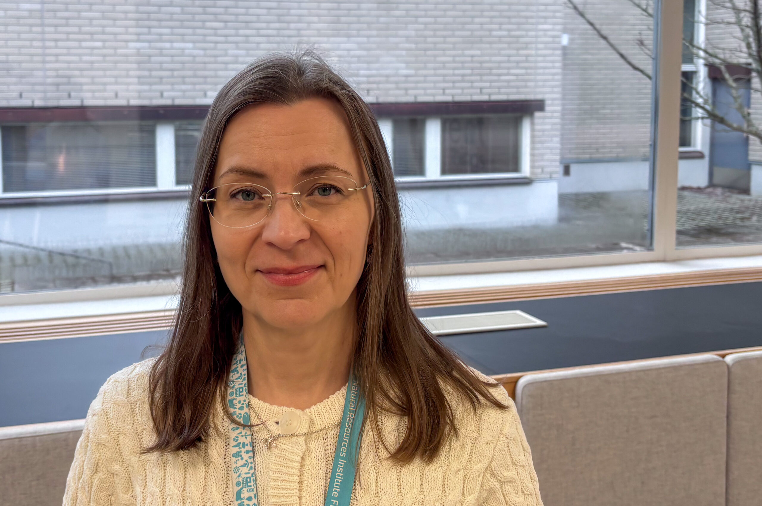
Eeva Vainio, Research Professor at the Natural Resources Institute Finland, hunts for viruses that infect wood-decay fungi and studies their properties. The goal: to use these diseases to control decay fungi and save the forestry industry tens of millions of euros.
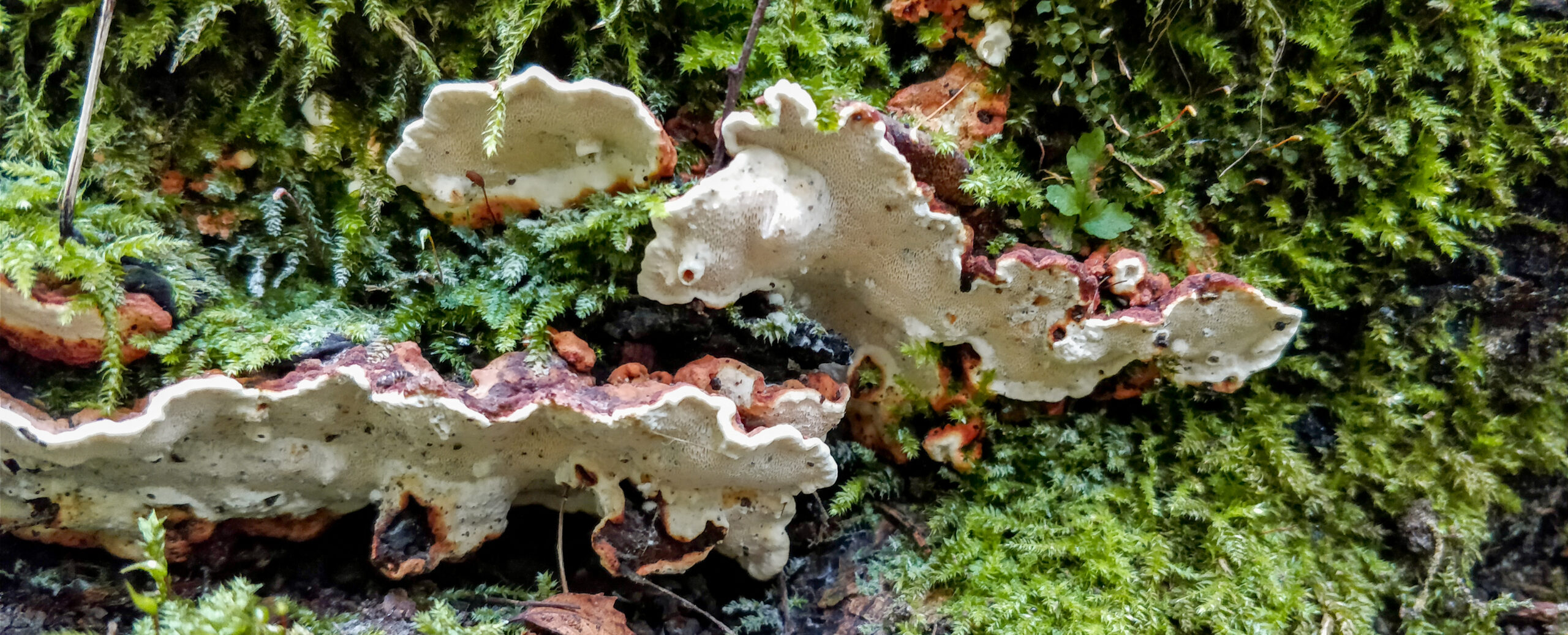
If you spot Research Professor Eeva Vainio out picking mushrooms, there’s no point trying to spy on her favourite foraging spots. In the forests of Espoo, Vainio isn’t looking for porcini or chanterelles. What interests this Natural Resources Institute researcher are root-rot fungi, honey fungus and other wood-decay species. And these are no minor organisms. The root-rot fungus Heterobasidion parviporum, for example, causes a disease in which the base and root system of a spruce tree can rot several metres up the trunk.
Wood-decay fungi rarely top anyone’s list of welcome finds. Besides being mostly inedible, they cost Finnish forestry tens of millions of euros a year. A single root-rot individual has been known to infect more than 50 trees. Honey fungus, meanwhile, sends long root-like threads through the soil, reaching from one tree to the next.
“For a researcher, wood-decay fungi are far more fascinating than the edible kind,” Vainio insists.
But fungi, captivating as they are, aren’t actually Vainio’s main focus. Her real interest lies inside them. Wild fungi harbour a rich variety of specialised viruses. Vainio hopes that one day these pathogens can be turned against the very decay fungi they inhabit.
Much about fungal viruses remains unknown. One thing, however, is certain: there are a great many of them. As detection techniques have improved, new viruses keep turning up. According to Vainio, almost every fungus examined so far has been found to carry viruses.
“Not necessarily every single individual, but nearly all the species we’ve studied have them,” Vainio says.
“And as our knowledge grows, it’s quite possible that fungi previously declared virus-free would turn out to harbour viruses too, if we analysed them again with today’s methods.”
Unlike animal viruses, fungal viruses rarely cause aggressive, fatal diseases in their hosts. Fungal viruses simply don’t spread very efficiently: transmission from one fungus to another typically requires physical contact or some kind of external carrier, such as an insect that picks up the virus from an infected cell and delivers it to a new individual. A virus that kills its host quickly, then, doesn’t have great prospects of its own.
Even within a single individual, a virus may not spread all that effectively. A root-rot fungus that covers an entire hectare, for instance, might have parts where infection stunts growth right alongside perfectly healthy sections.
“You can have an individual where half is infected and half isn’t. And yet it goes on living like that for decades or even centuries.”
Still, Vainio says she sometimes marvels at the sheer number of viruses.
“You’d think it would interfere with the cell’s functioning.”
Finding viruses in fungi is no easy task, because infection often leaves no visible trace. Viruses are also far too small to see under an ordinary microscope.
To find them, you have to look inside the cell. The most effective approach is to examine the RNA contained in a fungal cell. RNA can be thought of as a working copy of the genetic information stored in DNA.
Unlike DNA, which contains the entire genome, a single strand of RNA covers just one gene. It carries that gene’s instructions from the DNA onward, telling the cell which protein to produce.
Not all RNA in a cell necessarily comes from the cell’s own DNA, however. If a virus is present, it produces RNA of its own in an attempt to hijack the cell into making copies of it. When viral RNA is detected in a cell, the virus itself can be identified.
This amounts to assembling quite a jigsaw puzzle. An RNA analysis begins by breaking open the cell and collecting all the RNA molecules inside it.
Unfortunately, the process also chops that RNA into fragments roughly 100 base pairs long. From this mass, ribosomal RNA — responsible for the structure of the RNA — is identified and filtered out. What remains is around 100 million fragments of about a hundred base pairs each. Researchers then try to computationally reassemble these into intact sequences a few thousand bases long.
Putting together a puzzle like that is impossible without supercomputers.
Reassembling the RNA from a single fungal cell takes trillions of calculations. A supercomputer can do it in a matter of hours.
The use of supercomputers in fungal virus research is fairly recent. As a government agency, the Natural Resources Institute Finland gained affordable access to one only in 2020. It has, however, revolutionised virus discovery: new ones keep appearing.
“For a biologist, it’s always exciting to keep finding things that are new and unknown to science,” Vainio says.
“It’s a bit like going on an expedition without having to travel to the Amazon.”
Her research group has discovered dozens of previously unknown viruses.
Discovering viruses is only the beginning, though. After that, Vainio and her colleagues try to work out how the viruses spread and how they affect fungal behaviour.
One key question, for example, is whether a virus spreads via fungal spores and how effectively it transmits vegetatively to other fungal individuals.
In their analyses, Vainio and her team have also used Chipster, a software platform developed by CSC. It can be used, for example, to compare differences in gene expression between virus-infected and virus-free parts of the same fungal individual.
Vainio speaks highly of CSC’s services.
“The Chipster training course I attended years ago, for instance, was excellent.”
The search for fungal viruses is not purely basic research. The idea behind these projects is to find viruses that could be used to control root-rot fungi and other wood-decay species that cause major damage.
So is the plan to wipe out decay fungi with a pandemic?
“I’d say the aim is to prevent root rot and other fungal diseases from spreading at the scale they do today,” Vainio replies.
Fungal viruses generally spread too weakly for the word “pandemic” to apply. They don’t appear to be particularly lethal, either.
“But a virus that could stop an existing disease pocket from spreading or releasing spores would already be a good, natural control method. It could limit the disease from passing on to the next generation of trees.”
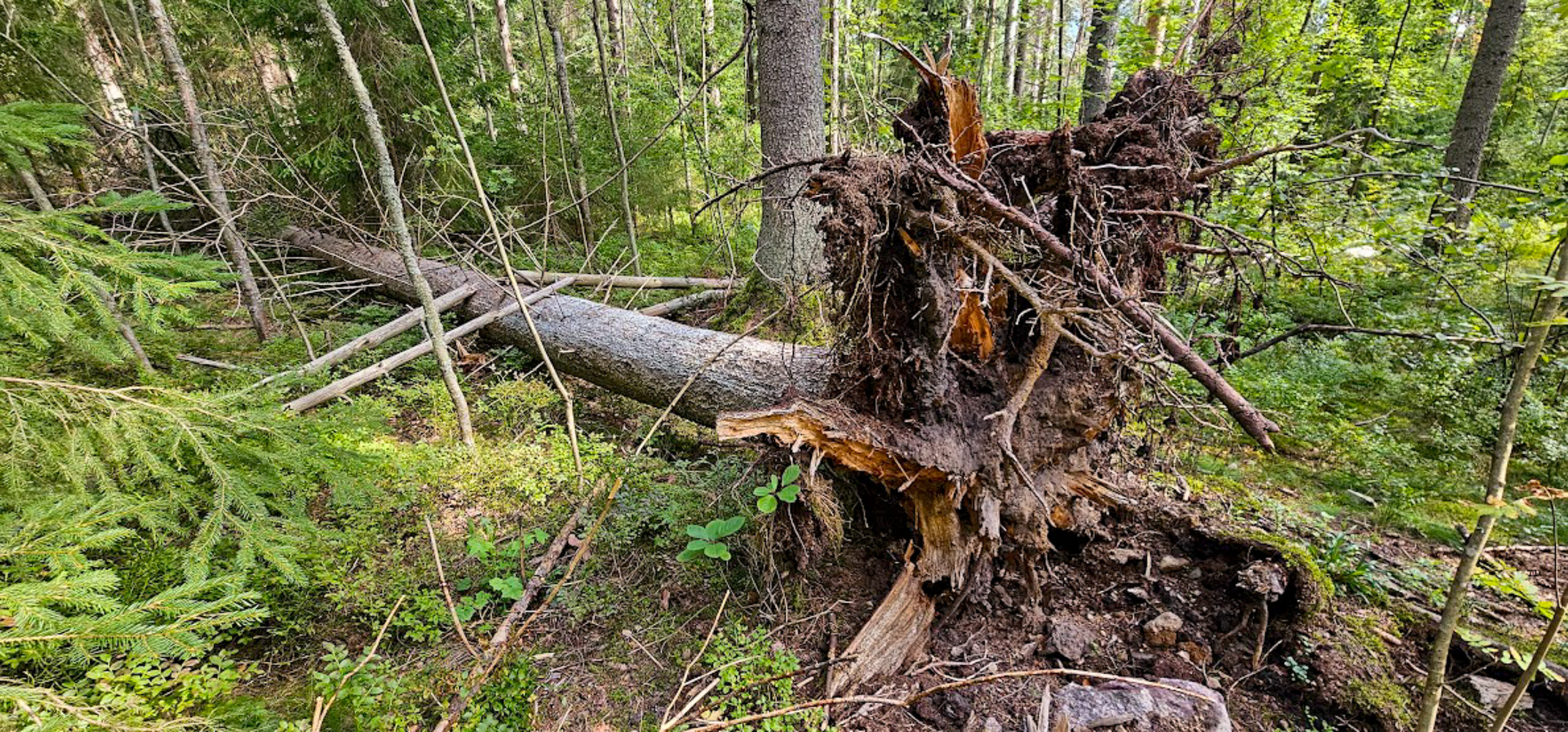
Using viruses to combat wild fungi may sound like a drastic intervention in the cycles of nature. In reality, however, the current spread of root rot is itself largely a consequence of human activity.
A clear-cut carried out in summer, leaving above-ground stumps behind, creates ideal conditions for decay fungi to spread. A fungus growing in a stump can propagate via both spores and underground root-like threads to nearby seedlings, which in a commercial forest are often the same species.
“In biodiverse natural forests, root rot has far fewer opportunities to spread,” Vainio points out.
“The stumps are surrounded by broadleaf trees that are less suitable as food for root rot, and the competing fungal communities are more diverse.”
Root rot became more common in step with the rise of summer felling. Treatment programmes introduced in the 1990s blocked spore-borne infections but did nothing to stop the fungus spreading through root systems.
Using viruses as a control measure, then, would be less about disrupting nature and more about restoring a more natural state of affairs.
Text: Juha Merimaa
Photos: Juha Merimaa and Eeva Vainio/Natural Resources Institute Finland
17 June 2026
Read the article in PDF format.


 ELIXIR is partly funded by the European Commission