
Bioinformatics has been used to crack the human genome. New imaging techniques will now provide us with a direct view of how genes interact with each other and their environment.
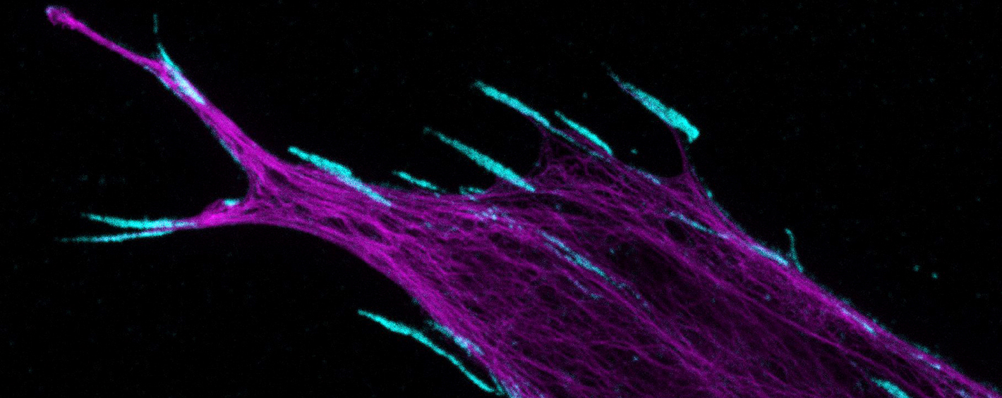
Modern imaging methods can be used to obtain accurate structural images of the body. They can be used to diagnose diseases, and plan treatments and monitor their effects. Imaging has taken huge leaps ahead. Nowadays, we can examine and analyse even living cells to the accuracy of individual molecules.
Turku BioImaging (TBI), provided jointly by the University of Turku and Åbo Akademi University, offers top-quality imaging technology to researchers. TBI also trains researchers in the use of modern biomedical imaging techniques and develops international infrastructures in the field. Computer modelling and software development is needed for the processing and analysis of image data.
“Some of the methods were already invented in the 1950s, but technology enabling their use by researchers took a long time to arrive. Now, the situation is different. Use of these methods has simply snowballed. This has been made possible by better lasers and computers, and the discovery of certain self-illuminating molecules and super-resolution techniques. Thanks to these advanced methods and techniques, we can see things that used to be in the realm of science fiction,” says TBI’s Administrative Director Pasi Kankaanpää.
Various techniques are used in imaging. Turku offers at least the following imaging services: light microscopy, electron microscopy, atomic force microscopy, positron emission tomography (PET) and magnetic resonance imaging (MRI). Turku also offers facilities for analysing thousands of cells and their properties on the basis of flow cytometry. Plenty of open-source software is available for analysing any data obtained.
“Imaging is important for research. You can comfortably say that it is now one of the most important areas in biological and medical research of all kinds,” says Kankaanpää.
He refers to surveys carried out in Finland to determine key biomedical research methods and their use. Although bioinformatics has helped to determine many aspects of the human genome and other species, and biomedical research has made huge progress, Kankaanpää says that this is not enough. Research also requires imaging.
“Now we need to find out what genes do and how they interact with other genes and their environment. What would be a better way to do this than simply seeing what is going on.”
According to Kankaanpää, image material in itself is not enough to guarantee results. In recent years, a new field of science, known as bioimage informatics, has emerged. It means methods that are used to manage images and specifically analyse them quantitatively. The size of a single three-dimensional model can run up to several gigabytes. A huge amount of image data is processed to extract genuine information and to understand what is going on in the images. Analysis can also be automated through machine learning, for instance.
“Bioimage informatics has been forecast to have the same kind of revolutionary potential as gene technology had a few decades ago. Now we can use imaging to analyse the birth of illnesses and the operation of cells.”
TBI offers, for example, cell imaging services that utilise the best light microscopy available. Such equipment can be used to take images of individual molecules or small living creatures in their entirety.

Microscopy is based on the wave movement of light or electrons. As the name implies, the light source of electron microscopes is a ray of electron particles with which the sample is bombarded. An electron microscope has a much better resolution than a light microscope. The resolution is up to thousands of times better, capable of an accuracy of 0.2 nanometres. Although on electron microscope enables us to obtain images of structures and cell organelles, it cannot be used on living cells, because the sample preparation will destroy the sample in practice.
The Turku unit has hosted some major achievements in terms of microscopy. Stefan Hell, who used to work at the Biophysics Laboratory of the University of Turku, received the Nobel prize in chemistry in 2014 together with Eric Betzig and William Moerner for the development of extremely accurate light microscopy. He conducted the crucial experiments in Turku in 1993–1996.
Light waves cannot directly create as high a resolution as an electron microscope can, but you can get around this with clever use of lasers and fluorescent molecules. These methods make use of fluorescence, that is, a molecule’s ability to absorb light at certain wavelengths and to send back light at a higher wavelength.
The fluorescent protein, or fluorophore, is attached to the molecule being studied within the cell for instance by means of genetic engineering or antibodies. In a manner of speaking, the fluorophore is used to “dye” the object being studied.
By using fluorescent markers by, so to speak, switching the light “on” or “off” in various ways, the latest light microscopes can see structures that were once only visible to electron microscopes. One such method is stimulated emission depletion (STED) microscopy. This can reach an accuracy of just of few nanometres, that is, millionths of a millimetre. The wavelengths of visible light are several hundred nanometres.
STED microscopes can show the cell organelle structures and even individual molecules and their functions in tissue. STED microscopes can also create three-dimensional image data, and can be used for living samples.
“We have used advanced light microscopes to examine flu viruses to see how they invade a cell. The analysis software we have developed has enabled us to calculate the percentage of the viruses that has entered the host cell and how many have remained outside. We can also follow where the viruses go within a cell, how fast they move and when they break down.”
Imaging has been used as a model to develop nanoparticles that can deliver drugs with precision inside a cell, by imitating the operating mechanism of the viruses. In cancer treatment, for example, small particles have been inserted into a metastasis with a catheter to target radiation treatment at a tumour.
“We can now obtain 3D image data of living cancer cells and see how the particles move. In the same way as with flu viruses, we imaged how particles enter a cell and how they break down.”
The objective is to obtain a silver bullet that does not affect healthy cells.
“Our goal is to target the drug directly at the cancer we want to kill and no other cells. This would reduce the side effects of cancer drugs. Imaging enables this type of development work. It is extremely difficult to imagine how this type of work could be done without modern imaging,” says Pasi Kankaanpää.
Ari Turunen
28.2.2019
Read article in PDF
Citation
Ari Turunen, Pasi Kankaanpää, & Tommi Nyrönen. (2019). Imaging helps to highlight significance of data. https://doi.org/10.5281/zenodo.8118822
More information:
Turku BioImaging
Euro BioImaging
https://www.eurobioimaging-interim.eu
CSC – IT Center for Science
CSC – The Finnish IT Center For Science is a non-profit, state-owned company administered by the Ministry of Education and Culture. CSC maintains and develops the state-owned, centralised IT infrastructure.
http://www.csc.fi
https://research.csc.fi/cloud-computing
ELIXIR
ELIXIR builds infrastructure in support of the biological sector. It brings together the leading organisations of 21 European countries and the EMBL European Molecular Biology Laboratory to form a common infrastructure for biological information. CSC – IT Center for Science is the Finnish
centre within this infrastructure.
https://www.elixir-finland.org
http://www.elixir-europe.org


 ELIXIR is partly funded by the European Commission