Brain researcher Jetro J. Tuulari is interested in nearly everything that shapes a child’s developing brain. To explore these questions, he relies on large datasets and substantial computing power.
A small boy experiences maltreatment in childhood. He becomes anxious and stressed.
Decades later, the boy becomes a father. Although his later life has been calmer, the stress of childhood still might have left an intergenerational imprint. In a study, Finnish researchers found a positive association between paternal maltreatment experiences and fractional anisotropy of their newborn baby, which implies faster pace of organization within the white matter.
Could the father’s childhood stress have accelerated the development of the child’s brain? And what might that imply for later life of the neonate?
At first glance, a causal link between the father’s early experiences and the newborn’s brain may seem almost implausible. Genetics cannot explain it: the father’s genome was fixed long before his childhood experiences. The sperm cell that initiated the child developed long after those early adversities.
The mechanism remains unknown, as does the longer term implications, but the association was clear in our study and a follow up study that found associations between childhood maltreatment and sperm epigenome, says neuroscientist Jetro J. Tuulari. Based, at the University of Turku, he leads the FinnBrain Neuroimaging Lab, which is part of a large FinnBrain Birth cohort that investigates children’s brain development.
FinnBrain research was launched at the University of Turku in 2010, and its purpose is to study the combined influence of environmental and genetic factors on child development and later health outcomes. The interest of this research touches one of the central questions of human development: what matters more – genes or environment?
From that starting point, the research has expanded in many directions, including to fathers.
“I tend to branch out a bit in my research. For example, from brains MRIs to sperm cell epigenetics,” Tuulari says.
Research on intergenerational exposures in fathers is only one of Tuulari’s many interests. His work has two main pillars: brain imaging and large datasets.
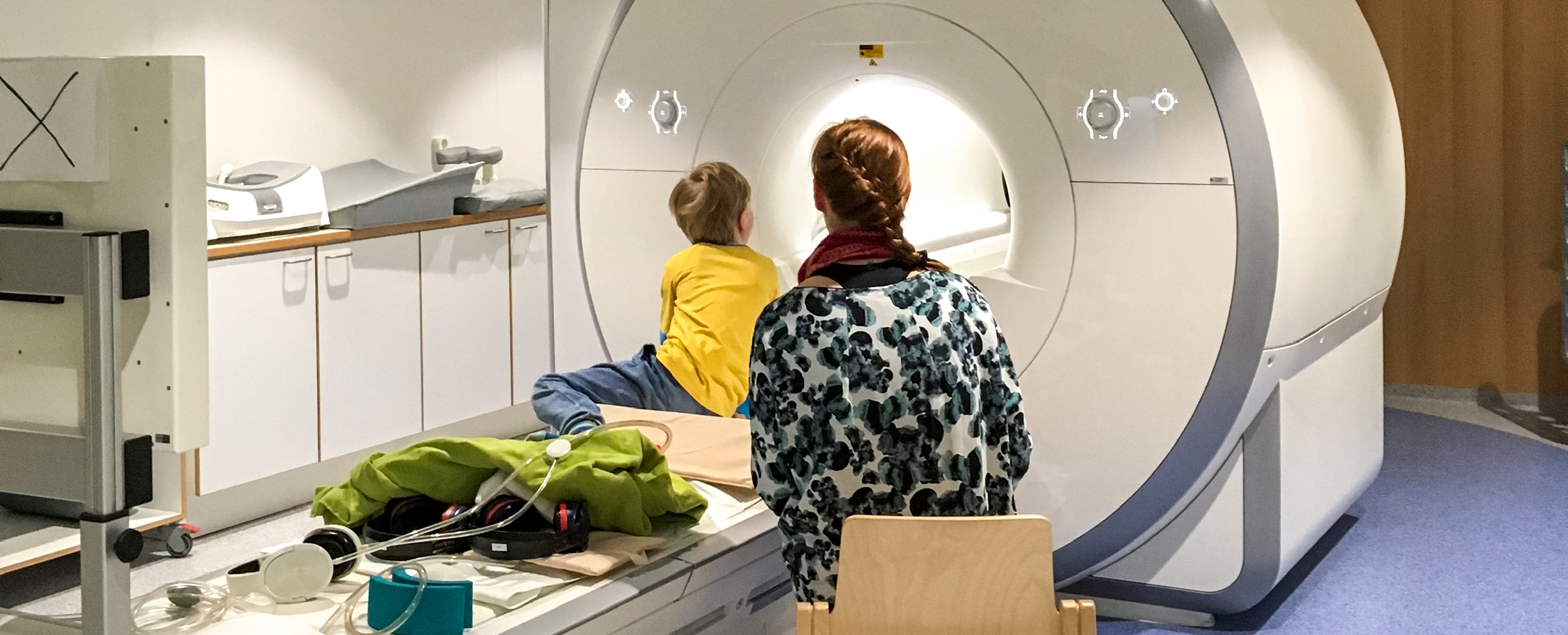
In FinnBrain, his primary research benefits from roughly 4,000 families enrolled in FinnBrain from Southwest Finland and Åland. We have scanned ca. 200 children with brain MR imaging at two weeks, five years and eleven years of age. The hope is to perform follow up imaging in the coming years, when participants are around 16 or 17, depending on funding.
In addition to FinnBrain data, Tuulari also uses large international datasets, such as the American Adolescent Brain Cognitive Development dataset, which includes biannual brain imaging of 12,000 individuals.
These data are large in scale, but he would prefer datasets that are even larger.
“I would like to move from datasets of thousands of people to datasets of tens of thousands.”
Achieving this requires harmonizing and combining brain imaging datasets collected worldwide. Artificial intelligence helps in solving these challenges by for instance enhancing image resolution based on existing models.
Large datasets make it possible to explore many research questions. Ever since his doctoral dissertation that focused on brain changes of obesity in adults, Tuulari has been interested in the relationship between children’s brain development and weight. In his latest study that is available as a preprint, he and his colleagues identified an intriguing correlation: the dynamic properties in well-established functional brain network at ages 9–10 appeared to be associated with current body weight and weight gain later in adolescence.
“Dynamic network properties do not explain the whole phenomenon as they capture around 20 per cent. Even so, that is clearly more than previous models using functional MRI data have found.”
The research does not yet explain why network dynamics influence weight gain. Possible hypotheses include differences in impulse control or in the sensation of hunger.
“Even so, it could be relevant, for example in identifying risk groups,” Tuulari says.
Another topic that has recently interested Tuulari is how brain size develops from birth to adulthood, which he has had the chance to study as part of the Lifespan Brain Chart Consortium that is led by Richard Bethlehem from the University of Cambridge and Jakob Seidlitz from the University of Pennsylvania.
“The idea is to define growth curves for the brain structure somewhat like the height/weight curves used in pediatric settings,” Tuulari explains.
“In other words, if a newborn’s brain is imaged, can we predict from that image how large the brain will be in young adulthood?”
The question is not simple. Brain growth in primary school age appears to involve growth spurts of different parts of the brain similar to the adolescent height spurt: if the brain grows rapidly in the pre-teen years, growth may stop earlier, whereas slower-growing brains may ultimately become larger.
But what would be the purpose of predicting brain growth? Would it be to identify those that are at risk for later life outcomes ?
“Not at this stage,” Tuulari says. Even the relationship between brain size health is not straightforward, apart from neurodegenerative diseases that are known to cause atrophy in the brain. In young children, for example, larger brains have been linked to an increased risk of autism so that bigger is not always better.
“Primarily, this is basic research at the moment. But like the growth charts used in child health clinics, brain charts have substantial potential for future clinical applications.”

Tuulari works with large neuroimaging datasets containing vast amounts of information. A typical dataset consists of three-dimensional brain images that contain vast amounts of information. Typically, tens or even hundreds to thousands of such images would be available for a study.
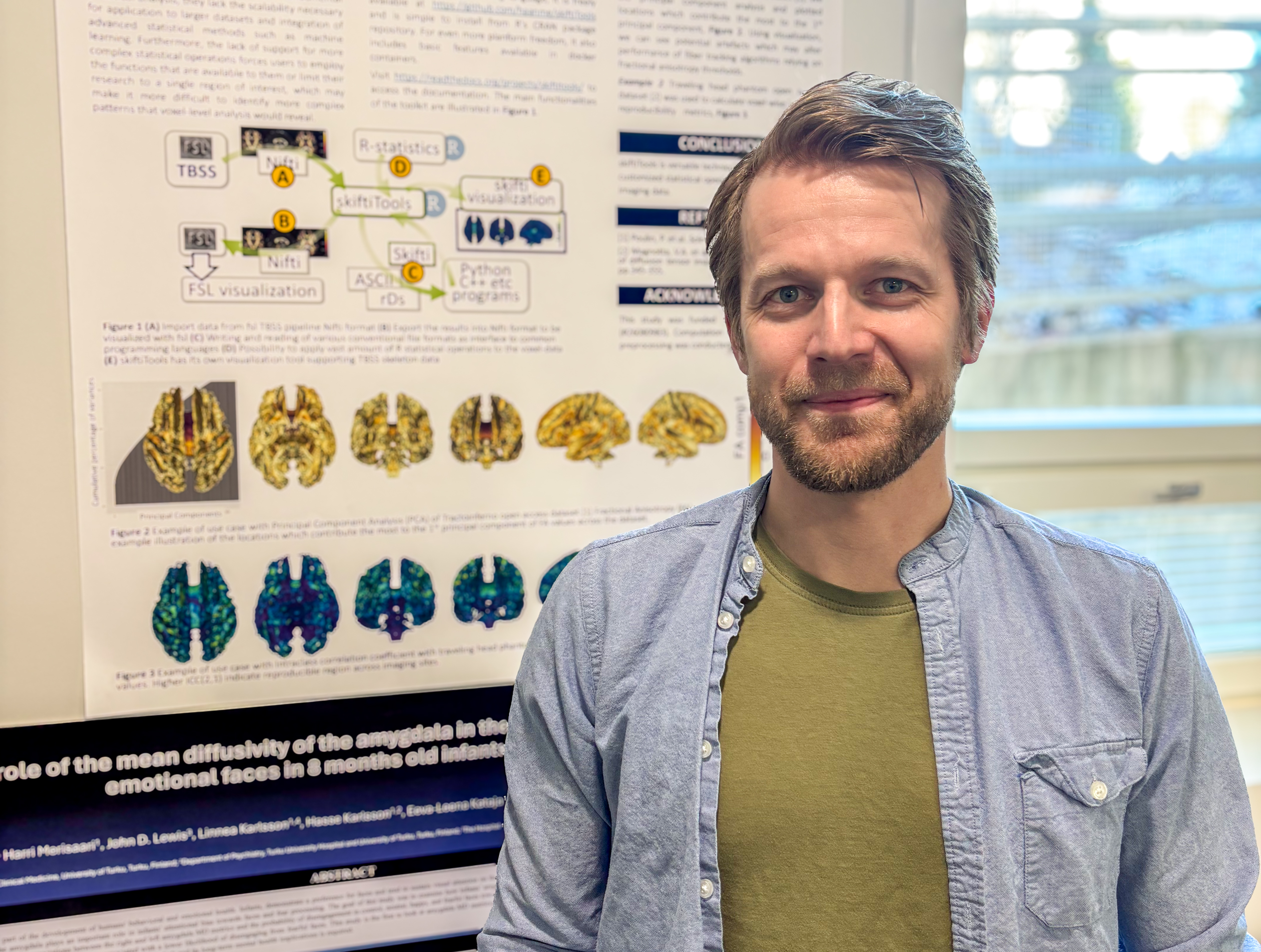
One of the most recent studies has focused on combining neuroimaging data formats with widely used statistical tools. Tuulari and Harri Merisaari, PhD in computer science developed a method for converting brain images into table format. One three-dimensional brain image becomes a table with about 192,000 entries. On the other hand, when we process functional MRI scans, images are taken every 1-2.5 seconds, and a session may last up to ten minutes. One series therefore might produce around millions of data points.
When thousands upon thousands of brain images are compared, the number of data points increases rapidly. Supercomputers are therefore needed for the data processing and statistical modeling.
Tuulari uses both the University of Cambridge supercomputer and CSC – IT Center for Science’s Puhti supercomputer. Each has its advantages.
“We have found the CSC services highly flexible and suitable for our tailored software and tools. “
But what about the biggest question of all? Based on vast brain imaging datasets, a slightly more than a decade of experience in studying the developing brain, what conclusion does Tuulari draw? Which is more relevant for development: genes or environment?
The research shows clear evidence of both. Genetics influences a child’s development, but findings such as the effect of a father’s childhood maltreatment on the child’s brain show that environmental influences may influence development in subtle ways and even extend across generations.
Even so, Tuulari leans toward genetics during early life.
“Just think about how complex an organ the brain is. When a foetus develops, the entire programme for building the brain is encoded in the genome and chains of amino acids forming proteins that are used to build a completely new individual. It is a true miracle.”
“The genome contains the instructions for what the structure of the hand is, as well as the blueprint for functional brain tissue. That is why, in my view, genes are more important during the first phases in a child’s development, and perhaps lifestyle and other factors would have more relevance during later life.”
Text, Jetro Tuulari’s photograph and video: Juha Merimaa
10.4.2026
Read the article in PDF format
More information:
FinnBrain
The University of Turku’s FinnBrain Birth Cohort Study aims to assess the role that environmental and hereditary factors play in the development of children. The study involves over 4,000 families with children who are monitored from the onset of pregnancy until late in adulthood.
Read more about children’s MRI studies on the FinnBrain research blog: https://sites.utu.fi/finnbrain/iltaunia-ja-seikkailua-lasten-ehdoilla-aivojen-mysteerien-jaljilla/
CSC – IT Center for Science Ltd
is a non-profit limited company owned by the state and administered by the Ministry of Education and Culture. CSC maintains and develops the state-owned centralised information technology infrastructure.
http://www.csc.fi/
https://research.csc.fi/cloud-computing
ELIXIR
is a distributed European infrastructure serving life science research. It offers, in a combined way, data resources, software tools, training, cloud services and high-performance computing resources from 23 countries and the European Molecular Biology Laboratory EMBL. Its Finnish node is CSC – IT Center for Science Ltd.
https://www.elixir-finland.org/
http://www.elixir-europe.org/


 ELIXIR is partly funded by the European Commission