


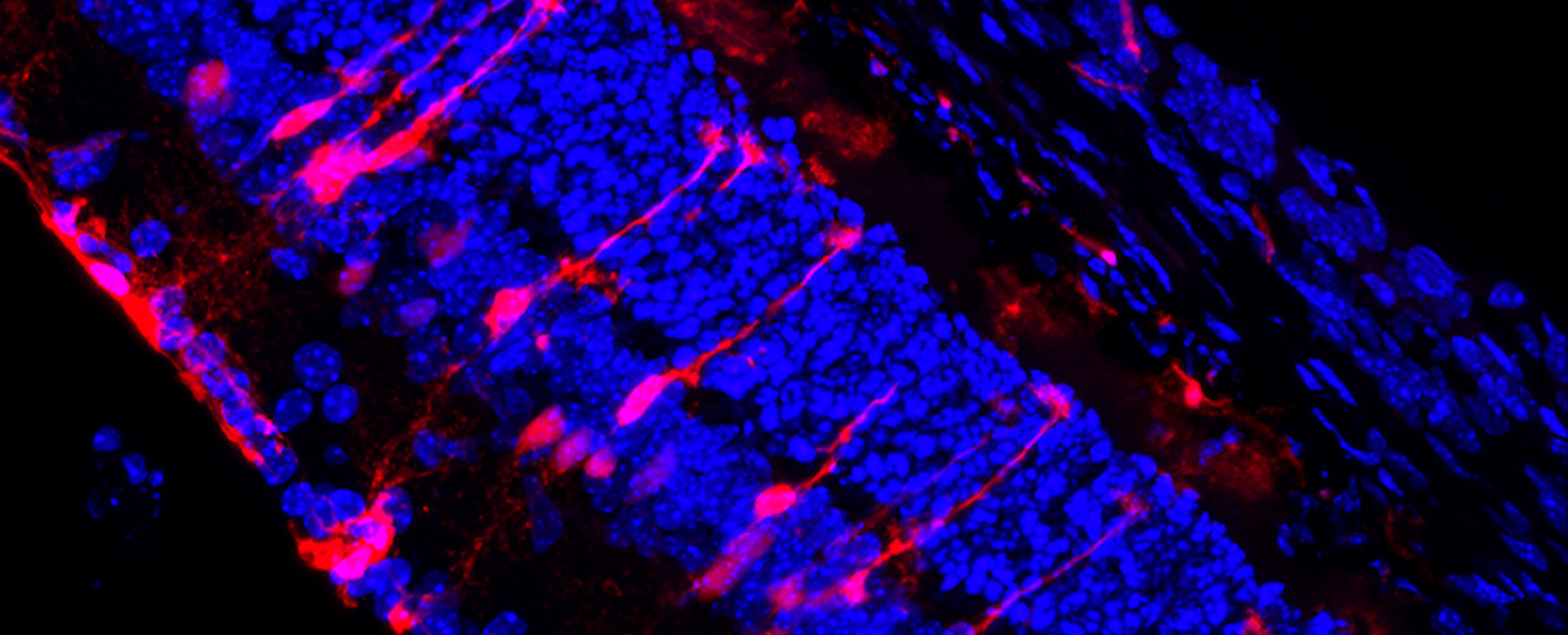
Fluorescence image of a mouse retina. The blue colour shows the retinal nerve cell layers, while the red colour highlights glial cells, whose role in mitochondrial diseases is being studied.
The same genetic variant may manifest as epilepsy in one person and Parkinson’s disease in another. Together with her research group, Academy Professor Anu Wartiovaara is investigating why mitochondrial dysfunction appears so differently in different individuals – and how it might be influenced.
“The mitochondrion is the powerhouse of the cell.” Many people remember this phrase from secondary school biology lessons. It has even occasionally been used humorously as an example of how schools teach things that seem far removed from everyday life.
However, the importance of mitochondria is far from trivial, says Academy Professor Anu Wartiovaara.
“Mitochondria are the engine that drives our lives. Without mitochondria, life would most likely never have evolved beyond single-celled organisms.”
Mitochondria are distinct structures within cells, possessing their own DNA. They are believed to have evolved from single-celled bacteria that entered into a symbiotic relationship inside cells.
Within the cell, mitochondria regulate metabolism – in other words, how the cell either burns sugars and fats from food when nutrients are scarce, or repairs and builds its tissues when nutrients are available.
And this affects almost everything in the body. As a result, even minor errors in mitochondrial function can lead to severe metabolic diseases.
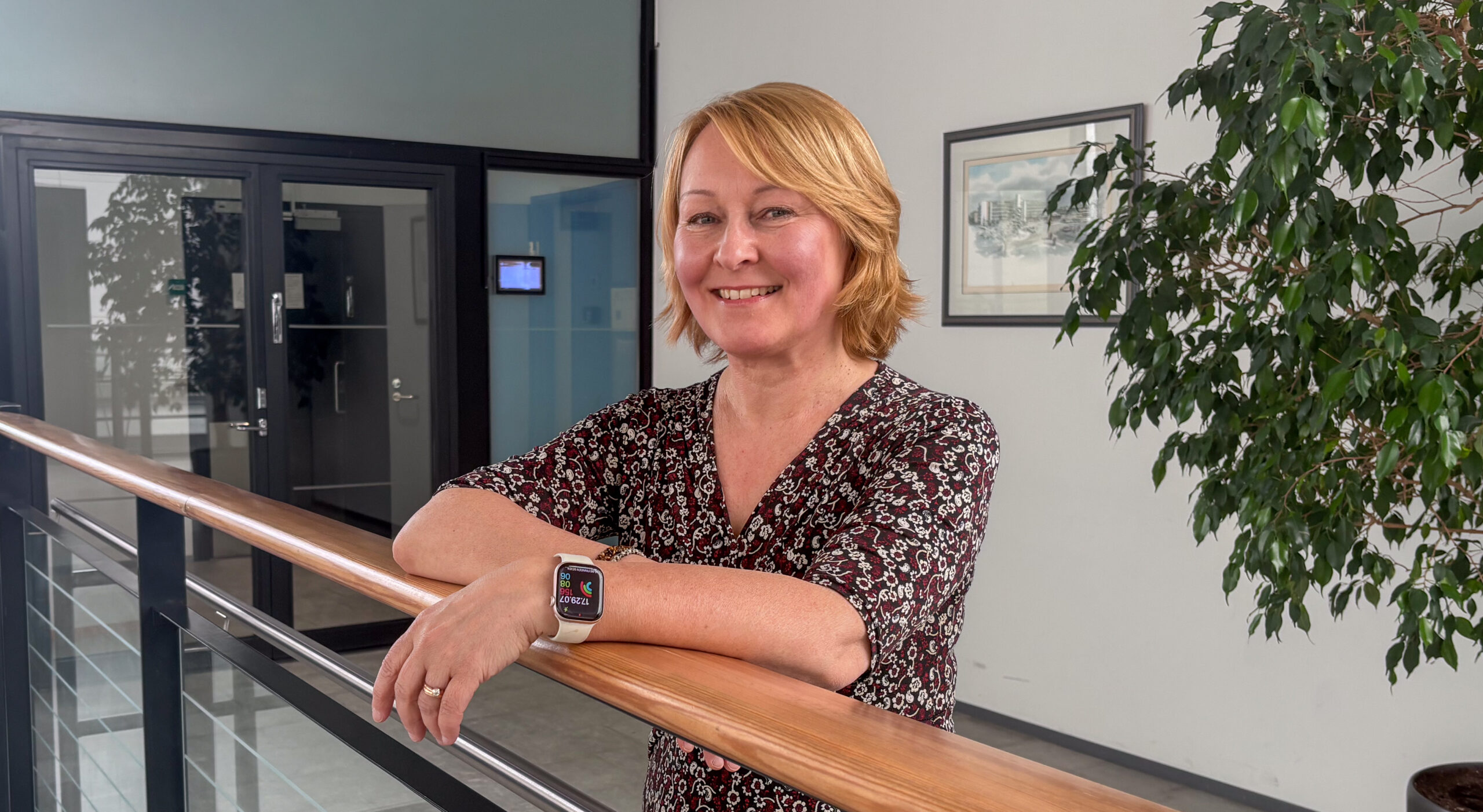
Mitochondrial diseases are serious, but they are also difficult to understand. Even when the disease-causing genetic variant is the same, it may manifest as Parkinson’s disease in one patient, epilepsy in another, and heart disease or muscle weakness in a third.
This is what interests Wartiovaara, Professor of Clinical Molecular Medicine at the University of Helsinki.
“Research on mitochondria has made us interested in why diseases manifest in certain tissues in the first place, but not in others.”
An intriguing example is MIRAS disease, discovered in 2005. This disease, inherited from a single founder mutation dating back to the Viking Age, affects how the body replicates mitochondrial DNA.
Approximately one in a hundred Finns carries the genetic variant that causes MIRAS, but the disease develops only if the variant is inherited from both parents.
The disease nevertheless appears in many forms: as teenage-onset epilepsy in one person, balance problems in adulthood in another, and Parkinson’s disease in middle age in a third. Because the genetic variant is the same, Wartiovaara’s group concluded that external factors must explain the different manifestations.
By testing potential factors, the group found that viral infections affecting the nervous system, such as herpes, tick-borne encephalitis and SARS-CoV-2, promoted the onset of the disease at a young age.
Because of the genetic alteration, cells fail to detect the virus in time, allowing it to multiply. This is followed by an overactive inflammatory response that particularly damages cells responsible for inhibiting activity in the nervous system.
“Our results seem to indicate that MIRAS-related epilepsy is associated with a reduction in cells that suppress neuronal activity. The clinical picture resembles viral encephalitis,” Wartiovaara explains.
“There is therefore probably a great deal in common between the mechanisms.”
Wartiovaara’s research spans the entire field of medicine, as mitochondrial diseases occur in every medical specialty. Sometimes the research is conducted using large patient databases, such as FinnGen, which includes data from 500,000 Finns.
Nevertheless, the primary focus of interest is metabolism at the molecular level – the underlying causes of disease. These are studied using a wide range of disease models, from cell cultures to flies and mice.
Research is also being advanced by rapid developments in measurement technologies: with current methods, biochemical reactions taking place within cells can be measured virtually in real time.
This generates enormous amounts of data. For example, a single tissue or blood sample can simultaneously yield more than a thousand different molecular markers.
Wartiovaara refers to this as a metabolic fingerprint.
“Each person’s fingerprint is slightly different, but among healthy individuals they resemble one another.”
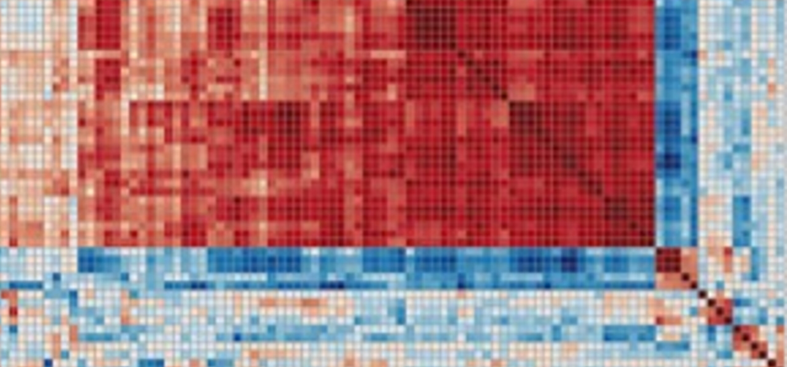
Even a small genetic defect can significantly alter the fingerprint. Wartiovaara compares the situation to a kaleidoscope. The thousands of metabolic products within a cell are like pieces of coloured glass in a kaleidoscope, arranging themselves into their own symmetrical patterns in each tissue. The pattern in muscle differs from that in the heart, and the pattern in the brain differs from that in the liver, and so on.
“When disease strikes, it disrupts this balance and the pattern rearranges itself into a new form that is no longer optimal for the function of that particular tissue.”
Together with her group, Wartiovaara is trying to understand the regulatory points that determine how the patterns in the kaleidoscope change.
“If we can identify the right regulatory molecules, we may be able to influence them and restore the overall metabolic system towards its proper form.”
Wartiovaara’s multidisciplinary research group includes physicians, biologists and biochemists alike. In recent years, computer scientists have also taken on an increasingly central role.
Wartiovaara’s group makes use of high-performance computing in many ways. First, they use it to model protein structures and suitable molecules that bind to them, which could be used as biosensors to measure the movement of metabolic products within the body.
When the group wants to determine where a particular metabolic product travels within the body, they seek to model the type of protein to which the compound binds. They then screen vast open molecular libraries to identify other protein structures with similar binding sites.
The libraries operate according to the so-called FAIR principles*, making it easy to search them for potential molecules that could be used as sensors.
“An ideal sensor molecule does not affect the functioning of the body but binds to the molecule we are looking for and signals its presence, for example by producing light,” Wartiovaara explains.
Such molecules make it possible to study the functioning of living cells.
Finding these molecules requires not only extensive molecular databases but also the high-performance computing resources, such as SD Desktop, Puhti, Allas and ePouta, provided by CSC – IT Center for Science.
Substantial computing power is also needed to compare metabolic fingerprints. Once metabolic profiles have been established for rare diseases, machine learning could be used to compare the profiles of unknown diseases with them. This makes it possible to make broader use of knowledge gained from rare diseases.
“If, for example, some patients with Parkinson’s disease resemble a known mitochondrial disease that also manifests as Parkinson’s disease in terms of their metabolic fingerprint, this may help guide treatment for similar types of disease.”
High-performance computing is also needed for integrating and analysing multiple layers of biological data. When information on proteins, metabolic products and gene expression is obtained separately from a single tissue sample, multiple layers of data are generated, and the volume of data becomes enormous.
Wartiovaara’s group is testing ways to combine this volume of data using computational capacity, and even to predict how metabolism is likely to occur in a given tissue and how it changes as disease progresses.
Overall, developments in data science have revolutionised basic medical research over the past couple of decades, Wartiovaara says.
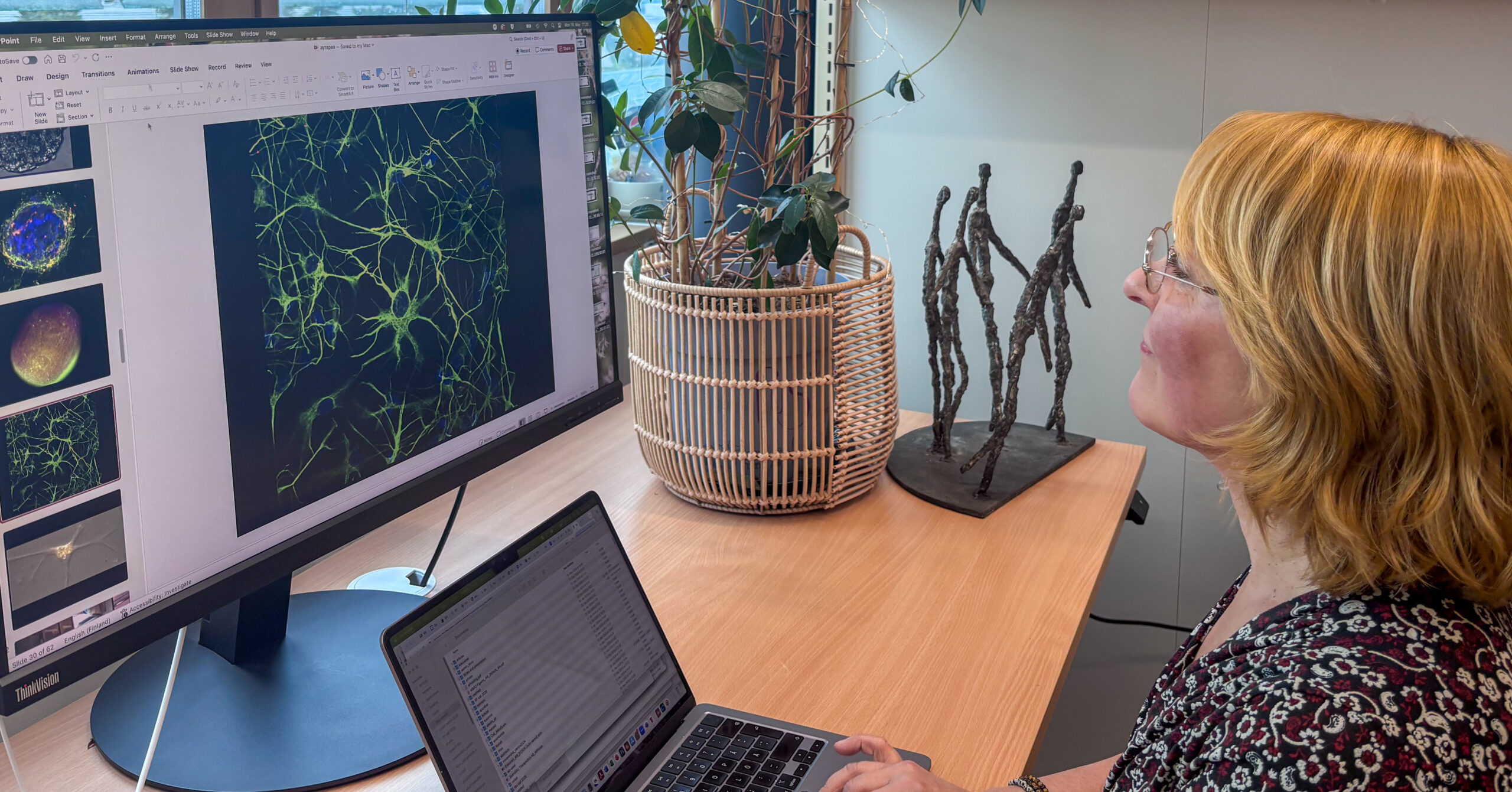
“Mitochondria convey signals about the availability of nutrients in our bodies and shape the growth and energy balance of our cells. The nature of these signals changes, either primarily or secondarily, in all diseases.”
The same genetic variant can manifest differently across patients . In addition, its manifestation may be influenced by nutrition, environmental toxins, viruses, or some combination of these factors. As a result, research into mitochondrial mechanisms generates enormous amounts of data and significant computational resources.
“Although we are constantly making progress, new questions continue to emerge all the time. That is what makes this so enormously fascinating.”
Text: Juha Merimaa
Photos: Juha Merimaa and Anu Wartiovaara’s research group
11 August 2026
Read the article in PDF format.
Further Information
FAIR DATA*
ELIXIR promotes the FAIR principles, according to which research data should be Findable, Accessible, Interoperable and Reusable.
Shared standards enable the efficient use of research datasets. For example, the UniProt database used by Wartiovaara contains sequence and annotation data on more than 120 million proteins.
https://faircookbook.elixir-europe.org
https://elixir-europe.org/what-we-offer/guidelines/data-management
CSC – IT Center for Science Ltd
CSC is a state-owned, non-profit limited company administered by the Ministry of Education and Culture. CSC maintains and develops Finland’s centrally owned scientific computing infrastructure.
csc.fi
research.csc.fi/cloud-computing
ELIXIR
ELIXIR is a distributed European life science research infrastructure. It brings together data resources, software tools, training, cloud services and high-performance computing resources from 23 countries and the European Molecular Biology Laboratory (EMBL). Its Finnish node is hosted by CSC – IT Center for Science Ltd.


 ELIXIR is partly funded by the European Commission