


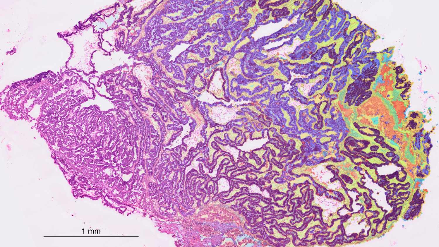
Visium HD dataset of a human ovarian cancer sample. The capture area measures 6.5 × 6.5 mm, containing approximately 11.2 million continuous capture spots. Dataset is publicly available from 10x Genomics; image edited by Franziska Niemeyer.
Spatial transcriptomics is such a new technique that it doesn’t even have established standards for analysis yet. University of Helsinki researcher Franziska Niemeyer learned the method on an international top-level course – and this spring brought it within reach of Finnish researchers too.
What is a cell up to? There are many ways to approach that question, but one of the most effective is to sequence the cell’s RNA. RNA shows which genes are active in the cell. From this, you can further deduce whether a cell belongs to, say, a tumour, or is healthy tissue.
The traditional way of doing sequencing has been to take a tissue sample and analyse it as one lump. A single sample, however, can contain millions of cells, and the final analysis ends up telling you more about what the sample’s cells did on average than about the activity of individual cells.
This kind of analysis could be compared to studying a Lego model by taking it apart and calculating averages of the pieces’ shapes, sizes and colours. This level of precision may be enough if, say, you just want to see whether a lump under the skin is a tumour – but sometimes more precise methods are needed.
A more sophisticated alternative is so-called single-cell analysis. To continue the Lego analogy: the model is taken apart further, but this time the pieces are sorted by type into some kind of order. This lets you focus on examining an individual piece. This kind of analysis, however, loses the bigger picture: even though a piece is red and has six studs, we no longer know where in the structure it was located.
When looking at cells, this is a problem, because the cells in the body don’t operate in a vacuum but interact with other cells. So, focusing on just one cell, without its relationship to neighbouring cells, never gives the full picture of what a cell is doing. Better techniques were needed.
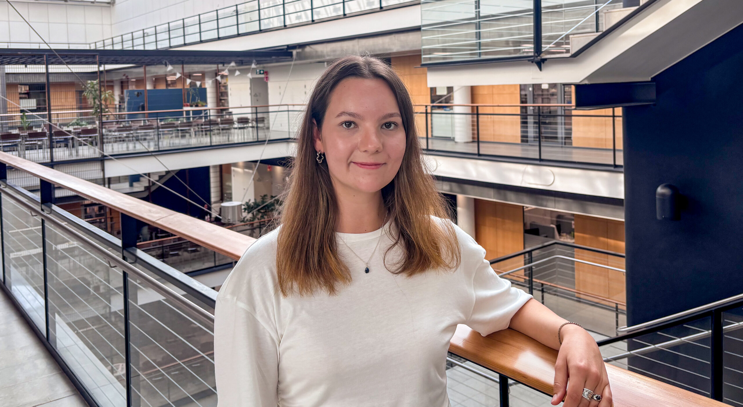
In 2018, Franziska Niemeyer began studying bioinformatics and genome research at Bielefeld University in Germany. Already at the start of her studies, she was interested in computer vision and image analysis – computers could analyse visual data more precisely and reliably than the human eye.
In 2022, during her master’s studies in Helsinki, Niemeyer was looking for a research group where she could pursue a PhD. She found Andrew Erickson’s newly established research group, which focused on prostate cancer and used a new and interesting technique: spatial transcriptomics. Niemeyer was eager to join the group.
It really was a brand-new technique. The key scientific breakthroughs behind spatial transcriptomics had only been made in 2016, and the first commercial applications came in 2019 – that is, after Niemeyer had already started her studies.
”The new technique seemed interesting right away. I wanted to learn more about it”, Niemeyer says.
The original sequence-based spatial transcriptomics is an analysis technique in which a tissue sample is placed on a special kind of microscopy slide with probes attached to it that can capture RNA molecules. It can be imagined as a piece of blotting paper onto which the tissue sample is placed and where RNA molecules released from the tissue are captured. At the same time, a high-resolution image of the tissue is taken. The output is essentially a map: for each position on the microscopy image of the tissue, you know which genes are active. The resolution of the technique varies, ranging from approximately 20 cells per capture location to subcellular resolution.
The technique has two challenges, however. First, it is new and therefore expensive: a single experiment with four 6.5 × 6.5-millimetre capture areas costs almost 15,000 euros.
Second, the technique produces enormous amounts of data. Even though a piece of paper a few millimetres across doesn’t sound like much, a single 6.5 × 6.5-millimetre sample capture area can hold as many as hundreds of thousands of cells. With hundreds of thousands of RNA molecules in each of them, each typically 1,000–2,000 bases long, the amount of data contained in a single sample climbs to levels that are too much for ordinary computers. There is also no established analysis workflow for processing this type of data yet.
”Getting to grips with analysing spatial transcriptomics data basically means learning to code your analysis”, Niemeyer explains.
The challenge didn’t scare Niemeyer off. In 2025, together with 30 other researchers, she attended a four-day spatial transcriptomics course organised by the ELIXIR Single-Cell Omics Community in Lausanne, Switzerland.
The course had lectures and analysis exercises. In these sessions, students used different types of analysis codes. Because the technology is still so new and every experiment is different, the code must be tailored separately for each experiment, in order to filter out what’s essential from the huge volume of data.
”The course was very inspiring and well organised. It was interesting to chat with some of the biggest names in the field over lunch”, Niemeyer recalls.
Still, the biggest surprise in the end was how unstructured the field still is.
”When you asked about standards in the field – for quality control, sample normalisation, or cell segmentation, for instance – you’d often get the answer that even the experts didn’t really know yet which method is the best.”
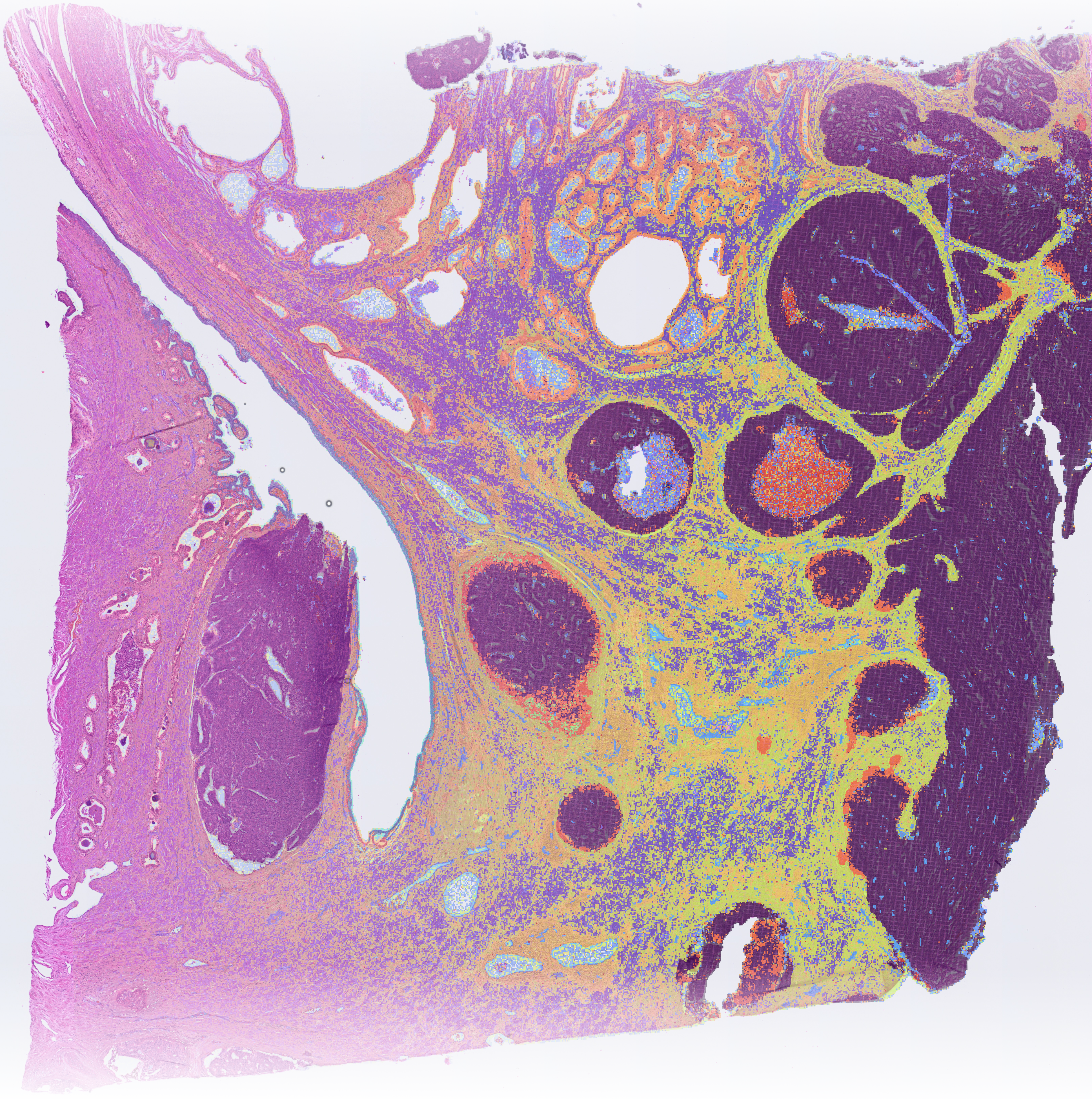
After the course, Niemeyer returned to Finland and began putting what she had learned to use in her doctoral research. It deals with the evolution of prostate cancer tumours and the development of non-invasive diagnostic tools.
”Lately my research has also turned toward developing open-source, automated workflows for spatial assays”, Niemeyer says.
She also decided to pass on what she had learned. In 2026, Niemeyer co-organised a spatial transcriptomics course at the University of Helsinki. Overall responsibility for the course lay with Associate Professor Anna Vähärautio, but Niemeyer handled the practical organisation and led part of the hands-on exercises.
The two-week course drew partly on materials from Lausanne.
”All the Lausanne lectures were recorded, so we could make them available for our course participants as well.”
Invaluable help also came from Noppe, the teaching platform run by CSC – IT Center for Science, where course participants could simulate the kind of computing normally done on a supercomputer.
”Noppe worked excellently. At first, we also considered booking time on an actual supercomputer, but for purely teaching purposes that wouldn’t have made sense. In the end, though, we gave instructions for using Puhti and later Roihu to those who wanted to make use of the technique in their own research.”
The Helsinki course also took in 30 participants, but unlike in Lausanne – where students were deliberately selected from across Europe – the majority of the Helsinki participants came from the University of Helsinki itself.
The course organised by the University of Helsinki represents the strength of the ELIXIR network, says Eija Korpelainen, Training Coordinator for ELIXIR Finland. She was one of the founders of the ELIXIR Single-Cell Omics Community and has been involved in its work from the start, since 2022.
She was also involved in selecting the participants for the Lausanne course. The idea was to spread knowledge and expertise as widely as possible, so participants were recruited from all over Europe.
”Training is essential for turning this expensive data into valuable knowledge.”
As a new research method, spatial transcriptomics is also developing rapidly, with commercial players bringing new technologies to market.
“The development of both laboratory technologies and analysis methods is so rapid that international collaboration is needed to identify the best approaches. By working together, we can also offer advanced courses for researchers.”
Niemeyer says she would be happy to help other universities organise similar courses in Finland.
Interest in spatial transcriptomics is strong across Finland and Europe. The latest international spatial transcriptomics course organised by the ELIXIR Single-Cell Omics Community took place in Uppsala on 6 October 2026. Eight Finnish researchers from different universities were selected to participate.
A project by the ELIXIR Single-Cell Omics Community has been selected for ELIXIR BioHackathon Europe 2026. In this project, researchers will develop methods for integrating single-cell datasets from different repositories and species to enable comparisons of corresponding cell types across species.
Text: Juha Merimaa
Photos: Juha Merimaa and Franziska Niemeyer
28 October 2026
Additional information:
ELIXIR Single-Cell Omics Community brings together European researchers and infrastructures working on single-cell and spatial omics. It develops shared standards, best practices and training to help the field’s fast-growing volumes of data become usable knowledge.
elixir-europe.org/communities/single-cell-omics
CSC – IT Center for Science Ltd
is a state-owned, non-profit company administered by the Ministry of Education and Culture. CSC maintains and develops centralised IT infrastructure.
csc.fi
research.csc.fi/cloud-computing
ELIXIR
is a distributed European research infrastructure for life science data. It brings together data resources, software tools, training, cloud services and high-performance computing resources from 23 countries and the European Molecular Biology Laboratory (EMBL). Its Finnish node is CSC – IT Center for Science Ltd.
elixir-finland.org
elixir-europe.org


 ELIXIR is partly funded by the European Commission